SINONASAL
POLYPOSIS
| เป็น soft tissue mass ในจมูกและ paranasal sinuses ที่ประกอบด้วย edematous hyperplastic mucoperiosteum ซึ่งสุมหรือกองเป็นรูปร่าง polypoid มีความแตกต่างจาก mucoperiosteal inflammation ของ chronic sinusitis ก็คือ มี mucous-secreting glands น้อยกว่าและมี disordered vascular bed นอกจากนี้ตัว polyp เองยังมี specific polysaccharide material ใน ground substance ทำให้มี fluid และ electrolyte ใน polyp ในปริมาณที่ผิดปกติ นั่นคือมี intercellular accumulation ของ fluid ทำให้ polyp โตได้ เนื่องจากว่าตัว polyp มี fluid accumulation ร่วมกับมี hypocellular nature ดังนั้น CT จึงเห็น polyp เป็น fluid density | ||||||||
| Sinonasal polyp ใน SSCT จะมีความแตกต่างจาก isolated sporadic polyps และ polypoid lesion อื่นๆ เช่น ethmoidal polypoid mucoceles, antrochoanal polyps และ sphenochoanal polyps | ||||||||
| Ethmoidal polypoid mucocele เป็น benign inflammatory condition โดย CT เห็นเป็น expanded opacified sinus และบางครั้งอาจมี contrast enhancing ethmoid complex อาจมี destruction intercellular septa ได้เล็กน้อย ethmoid polypoid mucocele แตกต่างจาก ethmoid mucocele คือ ethmoid mucocele ไม่มี internal architecture และไม่ enhance | ||||||||
| Antrochoanal polyp และ sphenochoanal polyps เป็น edematous hyperplastic submucosa ในผนังของ maxillary sinus และ sphenoid sinus และเจริญเติบโตผ่านทาง ostium ไปยังจมูก แม้ว่าจะมี histopathological correlation กับ mucous retention cysts แต่โดยส่วนใหญ่เชื่อว่าเป็น true polyps | ||||||||
| ในขณะนี้ยังไม่ทราบสาเหตุที่แท้จริงของ
sinonasal polyp แต่พบว่า มีความสัมพันธ์กันกับ atopic rhinitis (ทั้ง
allergic และ non allergic ), asthma, chronic sinus infection, cystic
fibrosis, aspirin intolerance และ Katagener's syndrome Cystic fibrosis พบ sinonasal polyp เป็น complication ใน children และ young adult ที่มี cystic fibrosis เกือบทั้งหมดจะมีโรคทาง sinus และ 10-20% ของผู้ป่วย cystic fibrosis มี nasal polyp โดยปกติใน children มักจะไม่พบ sinonasal polyp เพราะฉะนั้นถ้าพบต้อง screen หาโรค cystic fibrosis อาการของผู้ป่วย sinonasal polyp มาด้วย nasal stiffness 100% rhinorrhea 69% facial pain 60% headache 43% และอื่นๆเช่น anosmia, recurrent bout of purulent sinusitis ซึ่ง sinonasal polyp สามารถบรรเทาได้โดยใช้ intranasal steroids Sinonasal polyp เป็นคำที่ synonymous กับ chronic hypertrophic polypoid rhinosinusitis |
||||||||
| CT
IMAGING OF SINONASAL POLYPS SSCT เป็น imaging ที่ใช้ screen sinonasal polyp ได้แม้ว่าจะเป็นกรณีหลังผ่าตัด ยกเว้นต้องการแยกโรคออกจาก mucocele, nasal sinus-based neoplasm ซึ่งต้องใช้ axial image และ intravenous contrast medium ร่วมด้วย |
||||||||
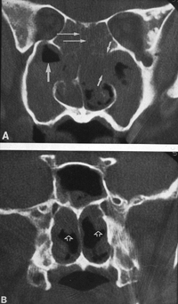
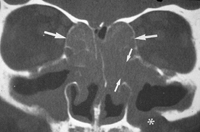
| Sinonasal polyp จะเห็นเป็น soft tissue density lesion โดยมีความแตกต่างจาก mucoperiosteal thickening โดยรูปร่างของมัน กล่าวคือมี polypoid shape ซึ่งพบ findings นี้ได้มากกว่า 90% มักพบที่ middle turbinate, lateral wall ของจมูก อาจพบที่ nasal septum ได้ sinonasal polyp มักจะพบทั้งสองข้าง imaging ที่พบมากเป็นอันดับสอง (89%) คือ infundibular enlargement แต่ต้องแยกโรคออกจาก antrochoanal polyp, ผู้ป่วยหลังได้รับการผ่าตัด และเนื้องอกใน sinus หรือ โพรงจมูก ส่วน imaging ของ sinonasal polyp อื่นๆที่พบได้ใน SSCT คือ polypoid involvement within the individual sinus, air-fluid level, bony attenuation, ethmoid sinus wall bulging | ||||||||
| Specific involvement ใน sinus โดย polyp จะให้การวินิจฉัยได้ยากเนื่องจากอาจเป็น sinonasal polyp ที่ secondary จาก obstructive inflammatory sinonasal disease และโดย process ของ sinonasal polyp มักจะเกิดในจมูกมากกว่าใน sinus พบว่า maxillary sinus และ ethmoid sinus เป็น sinus ที่มักพบ polypoid lesion ใน SSCT | ||||||||
| Air-fluid
level ใน paranasal sinus มีความสัมพันธ์กับ superimposed acute sinusitis
น้อยมาก พบใน sinonasal polyp ประมาณ 43% ต้องระวังอย่าสับสน bony attenuation หรือ deossification ของ nasal septum และ sinus trabeculae กับ partial volume effect แต่กระนั้นก็ตามถ้ามีการลดลงของจำนวน และขนาดของ trabeculae หรือ nasal septum ใน พื้นที่ที่ไม่ได้ทำการผ่าตัดมาก่อน ให้คิดถึงไว้ก่อนว่า อาจจะเกิดจาก aggressive inflammatory process เช่น sinonasal polyp โดย ethmoid trabecular attenuation พบ 63% ในผู้ป่วย sinonasal polyp และ nasal septum deossification พบใน sinonasal polyp 37% สำหรับการวินิจฉัยแยกโรคสำหรับ sinus และ nasal bony erosion คือ non malignant และ malignant กลุ่ม non malignant ได้แก่ dermoid cyst, mucocele, juvenile nasopharyngeal angiofibroma, inverting papilloma, pyocele, meningoencephalocele, Wegener's granulomatosis, fungal infection, syphilis, tuberculosis, sarcoid, cocaine nose ส่วน malignant ได้แก่ squamous cell carcinoma, rhabdomyosarcoma, lymphoma และ metastasis |
||||||||
| Bulging ของ lateral wall ของ ethmoid sinus ( lamina papyracea ) พบ 51% ของ sinonasal polyp โดยปกติแล้ว lateral ethmoid wall มักจะ straight หรือ convex medial configuration ถ้าพบมี sinus opacification ร่วมกับ obviously outward convex lateral ethmoid wall ให้สงสัยว่าจะมี sinonasal polyp โดยเฉพาะถ้ามี evidence อื่นของ sinonasal polyp ร่วมด้วย แต่ sign นี้ค่อนข้าง non specific อาจพบใน ethmoid mucocele และ sinonasal tumors | ||||||||
MRI
ช่วยบอก extension ของ sinus disease ได้ดีโดยเฉพาะในกรณีเข้าสู่ orbit
หรือ intracranially แต่ disease ของ sinus มักจะถูกจำกัดอยู่ใน paranasal
sinus และ nose MRI จะบอก รายละเอียดของ bone ได้น้อยกว่า CT และแพงกว่า
CT ( ภาพที่ 26, 27, 28 )
|
||||||||
| THERAPY
FOR SINONASAL POLYP การรักษาจะเริ่มต้นด้วยการให้ systemic steroids และ antibiotics โดย antibiotics อาจให้เป็น board spectrum antibiotics ซึ่งอาจจะให้ถึงหลาย courses ถ้ามีอาการบ่งถึงว่ามี infection อยู่ อาจต้องให้ antibiotic ต่อ 6-8 สัปดาห์ ทั้งนี้อาจให้ inhaled steroid ในการ maintenance FESS จะทำต่อเมื่อรักษาด้วยยาแล้วไม่ได้ผล |
||||||||
|
เมื่อวินิจฉัย
sinonasal polyp แล้วผู้ป่วยมักจะไปทำ screening radioallergosorbent
test with IgE level ถึงแม้ว่าการ desensitization ไม่เปลี่ยนแปลง sinonasal
pathology ก็ตาม บางครั้งอาจให้ antihistamine หรือ decongestant เป็นครั้งคราวและผู้ป่วยจะถูกแนะนำให้หลีกเลี่ยง aspirin การผ่าตัดจะทำในกรณี fail medical therapy, ในรายที่มี contraindication ต่อการให้ systemic steroids, complete nasal obstruction, uncontrol complication FESS ใช้ในกรณีผู้ป่วย sinonasal polyp เพื่อ resect polyp เพื่อให้เกิดอันตรายต่อ normal structure ให้น้อยที่สุด Common complication สำหรับการผ่าตัดคือ bleeding ทั้งจาก mucosal source และ จาก damage ethmoidal arteries Efficacy ของ surgical intervention ขึ้นอยู่กับ grade of severity ของ polyposis |
||||||||