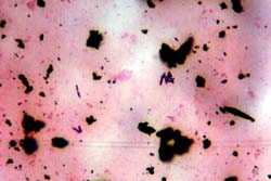
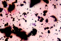
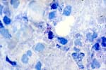
2. การตรวจจุลชีพจากสิ่งส่งตรวจ (Figure 4, 5, 6) ย้อมสีแกรม แยกยากจากกลุ่มของ Corynebacterium spp.ย้อมสี MAFB (Modified Acid Fast Bacilli) ให้ผลบวก เห็นรูปร่าง cocci หรือ
coccobacilli ติดสีแดง และบางเซลล์เป็น non acid fast อาจพบเป็นกิ่ง
4A
4B
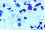
Figure 4 : Gram stain of pleural fluid in Amies transport medium (4A-4B) from
which were recovered Rhodococcus equi.
Note : The gram positive rod or coccobacilli difficult distinct from other gram
positive rod non-spore forming by gram stain.
5A
5B
5C
5D
5E
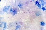
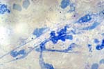
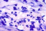
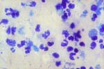
Figure 5 : Modified acid fast stain (MAFB) of pleural fluid from which were isolated Rhodococcus equi, illustrating rod or/and coccoid form with positive MAFB stain, characteristic of Rhodococcus. The MAFB may be stained on to part or fragment of the cells. (5A – 5E).
6A
6B
6C
6D
6E
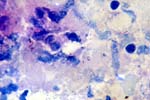
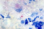
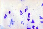
Figure 6 : Modified acid fast stain of sputum from which were recovered Rhodococcus equishowing short coccobacilli, some of which are modified acid fast stain positive (6A – 6F).
3. การเพาะแยก ขึ้นได้ดีบน blood agar ไม่ขึ้นบน Mac Conkey agar, สิ่งส่งตรวจที่มีเชื้ออื่นปนเปื้อนมาก เช่น เสมหะ ควรเพาะบน selective medium สำหรับแกรมบวก เช่น Columbia Colistin - Nalidixic acid agar, phenylethyl alcohol (PEA) blood agar